- Servicing the Triangle Area: Raleigh, Durham, Chapel Hill, Wake Forest, Cary, and Hillsborough NC

Common Eye Conditions In Cats And Dogs
Let’s start with a quick overview of terminology related to the eyeball to help you understand what you are seeing and reading.
- Ophthalmoscope - This is the tool our doctors use to examine the eyes. It uses lenses and illumination to better visualize the structures of the front and the back of the eye. Areas we are assessing with this tool are the pupil, lens, iris, optic nerve, retina, tapetum, blood vessels of the eye, cranial nerves, lash line, conjunctiva, cornea, and front and back fluid chambers of the eye.
- Globe - This is the term for the eyeball as a whole.
- Conjunctiva - The tissue on the inside of the outer eyelids and around the globe itself. This tissue can become swollen with trauma or infection.
- Cornea - The frontmost clear portion of the globe that allows light into the eye while protecting the iris and pupil. Since this area of the eye has no blood vessels (to keep it clear), injuries to this portion of the eye are difficult to spot without the help of a specialized fluorescent stain that sticks to any defects in this layer.
- Iris - This is the muscular colored circular portion of the eye that controls pupil size. This muscle can atrophy with age just like other muscle in the body and can develop small holes in it, especially in cats.
- Pupil - This is the hole in the middle of the iris through which light passes to the back of the eye. The pupil dilates (gets wider) in darker conditions to let more light in and constricts (gets smaller) in brighter light to let less light in. Humans and dogs have circular pupils, cats have vertical elliptical pupils, and horses and goats have horizontal pupils.
- Lens - This is a structure located behind the iris/pupil involved in focusing and adjusting light delivery to the retina.
- Retina - This is located on the back of the eye inside the globe and behind the iris and lens. This portion of the eye has cells that are activated by light signals and transmit information to the brain through the optic nerve. The retina contains rods and cones, blood vessels, and the macula and fovea (in humans and birds, but not cat and dogs).
- Optic nerve - This nerve transmits light information to the brain. It can be seen on an eye exam with an ophthalmoscope.
- Tapetum - This is a reflective membrane located just behind the retina that helps many animals see in low-light. Humans do not have this structure which is why we can’t see great in the dark compared to other species. This structure is responsible for the ‘glowing’ of animal eyes in a photograph and the color of this structure varies between purple, blue, green, and yellow depending on the animal’s eye color. Animals with 2 different colored eyes usually have 2 different colored tapetum.
- Eyelids - The outer layer of skin protecting the eyeball. While humans have 2 eyelids, cats and dogs actually have three. The outer eyelids can be voluntarily controlled when blinking. The 3rd eyelid, or innermost eyelid, is only raised passively when the globe is pulled back into the socket by a muscle behind the globe called the “retractor bulbi”.
- Retractor bulbi - This is a muscle that most mammals have (but not primates or humans) attached at the back of eye globe into the eye socket. When this muscle is activated, the globe is pulled deeper into the socket, causing the third eyelid to cover a portion of the globe. This is done to try and protect the eye from injury or to help shield the eye when painful.
- Eyelashes (cilia) - Small hairs located on the eyelid to help protect the eye. Humans have upper and lower eyelashes while cats and dogs only have upper lashes.
Common Eye Conditions in Cats and Dogs
There are many diseases of the eye in cats and dogs, but here is a list of some of the most common that we see in general practice. These conditions are all very different in cause and treatment, but you may notice that symptoms of these conditions are similar. This is why we always recommend having eye conditions looked at asap. Most of these conditions are painful or uncomfortable and some of these conditions can cause very serious lifelong complications (like blindness) if left untreated even just for a day or two.
Conjunctivitis
Inflammation and possibly infection of the conjunctiva (tissue inside the eyelid and around the eyeball). This finding can be secondary to allergy, trauma, bacterial infection, or viral infection.
Symptoms can include: Swelling or redness of the conjunctiva, eye discharge, squinting, raised third eyelid, and tearing. Sometimes we also see upper respiratory symptoms (sneezing, runny nose) with conjunctivitis and this can sometimes affect appetite.
Uveitis
Inflammation of the eyeball and the structures inside. In cats and dogs, this condition is often caused secondary to another systemic disease like Lyme infection, toxins, autoimmune diseases, cancers, or from blunt trauma to the eye area. We can also see uveitis as a result of other eye conditions that have gone untreated.
Symptoms can include: A cloudy or foggy appearance to the eye, iris color change, squinting, raised third eyelid, rubbing eye/face, lethargy, decreased appetite, light sensitivity, decreased vision.
Corneal Ulceration
This lesion is a wound/scratch to the frontmost clear portion of the eye and it is acutely painful. Typically a special fluorescent stain on the cornea is used to diagnose this condition.
Symptoms can include: Squinting, raised third eyelid, eye discharge, eye itching/rubbing, decreased energy, light sensitivity, constricted pupil.
Glaucoma
A very painful condition where there is a buildup of fluid pressure within the eye. This can be a primary disease or secondary to another underlying cause (like high blood pressure).
Symptoms can include: Redness of the eye, squinting, tearing, decreased energy or appetite, itching/rubbing of eye area, light sensitivity.
Cataract
A defect/cloudy area of the lens. Cataracts are often an incidental, age-related finding, but can also be secondary to other conditions like untreated uveitis and diabetes. If a cataract gets large enough it can significantly impact vision.
Symptoms are often not seen, but can cause decreased vision and can increase risk for other eye conditions like glaucoma and lens luxation.
Trauma
Any wound to or around the eye, but could also be blunt trauma to the bony structures around the eye. Failure to treat trauma can result in secondary conditions like glaucoma, uveitis, conjunctivitis, infection to the skin and soft tissue around the eye, vision impairment, or scarring. Remember, you can not see injury to the cornea with the naked eye- a special stain is required.
Symptoms can include: Wounds to skin around eye, swelling around eye, redness of conjunctiva, squinting, third eyelid raised, tearing or eye discharge, decreased energy or appetite.
Dry Eye (Keratoconjunctivitis Sicca - KCS)
A disease of decreased or defective tear production. Dry eye can be uncomfortable and can also cause blindness if left untreated. Certain breeds are prone to this condition like pugs, bulldogs, spaniels (cocker, CKCS, Springers), bostons, pekingese, samoyeds, shihtzus, westies, and yorkies.
Symptoms can include: Thick goopy eye discharge, squinting, scarring/‘browning’ of the cornea, raised third eyelid.
Signs Of Eye Pain In Animals Can Include:
- Decreased appetite
- Decreased energy
- Raised third eyelid
- Squinting or holding eye closed
- Constricted (small) pupil
- Excessive discharge from the eye
We recommend your pet be seen within 24 hours if you are seeing:
- Signs of eye pain (see above)
- Squinting, blinking, or holding the eye closed
- Thick or colored eye discharge
- Light sensitivity
We recommend your pet be evaluated immediately (at emergency hospital) if you see any of the following:
- Sudden change in color of the iris (colored ring-portion of the eye)
- Blood within the eye
- Traumatic wound or significant blunt trauma to the eyelid or eyeball
- Sudden blindness
- Cloudiness of the FRONT of the eye (note: cloudiness to the back of the eye is not an emergency)
Picture 1-3 (examples of various conditions that should be evaluated same-day): can not see the iris, can not see a portion of the iris, or iris appears hazy.
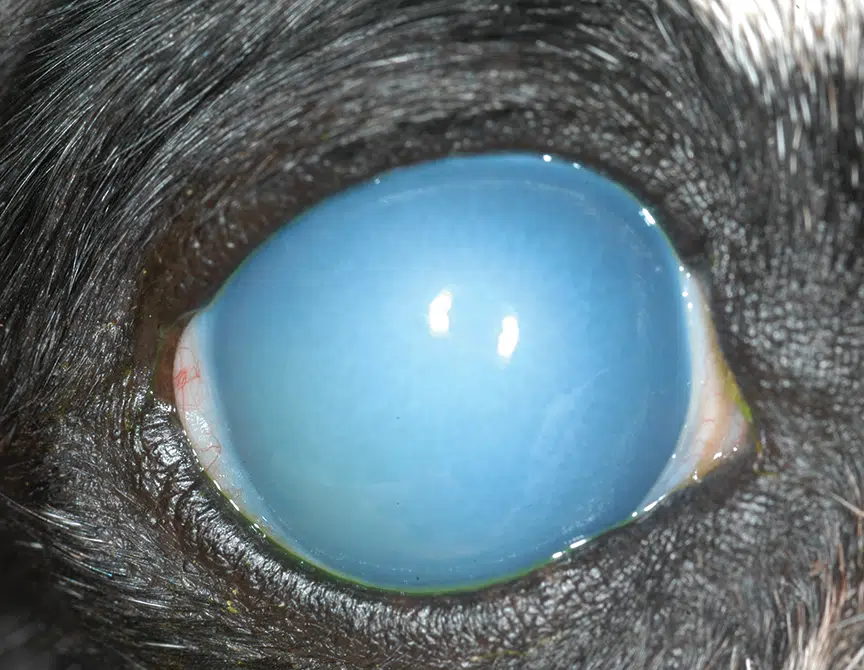
.png)
.png)
Photos from: Today’s veterinary practice, Science Direct, Vetlexicon
Picture 4 (not an emergency): iris is fully visible and clear and area behind iris is ‘milky’ or cloudy. This should be evaluated, but is not an emergency.
.png) Photos from: Today’s veterinary practice, Science Direct, Vetlexicon
Photos from: Today’s veterinary practice, Science Direct, Vetlexicon